The muscles of the thigh and lower back work together to keep the hip stable, aligned and moving. How pain in the upper thigh presents itself. It is part of the lower limb. Anatomy of the human body. This section of the website will explain large and minute details of arterial anatomy of upper legs (thigh arteries).
Thigh Muscle Images, Stock Photos & Vectors | Shutterstock from image.shutterstock.com How pain in the upper thigh presents itself. Anatomically, it is part of the lower limb. Thigh muscle anatomy hip anatomy gross anatomy human body anatomy yoga anatomy human anatomy and physiology anatomy study anatomy reference leg muscles anatomy. The probe is placed on the anteromedial aspect of the thigh, first in the short axis of the adductor longus, and then rotated into its. Muscles of the anterior thigh. The muscles and fasciæ of the thigh. The muscles of the thigh and lower back work together to keep the hip stable, aligned and moving. Top suggestions for upper thigh anatomy.
The probe is placed on the anteromedial aspect of the thigh, first in the short axis of the adductor longus, and then rotated into its.
630 anatomical structures of the upper limb (pectoral girdle, shoulder, arm, elbow, forearm, wrist we used the terminologia anatomica to label all the anatomical structures; We think this is the most useful anatomy picture that you need. Muscle anatomy lecture 12 photos of the muscle anatomy lecture anatomy muscles lecture test, muscle anatomy lecture, skeletal muscle. Mri of upper leg (femur). Top suggestions for upper thigh anatomy. The thigh is the area between the hip and the knee joint. The single bone in the thigh is called the femur. This webpage presents the anatomical structures found on thigh mri. Anatomynote.com found upper thigh muscle anatomy from plenty of anatomical pictures on the internet. Upper part of medial surface of the shaft of tibia. These images are from the visible human project sponsored by the national library of medicine. Anatomy of the human body. Anatomically speaking, the thigh refers to the region of your upper leg between your knee and your hip joint.
It passes obliquely across the upper and anterior part of the thigh, from the lateral to the medial side of the limb, then. In human anatomy, the thigh is the area between the hip (pelvis) and the knee. See more ideas about leg anatomy, anatomy, anatomy drawing. Mri of upper leg (femur). The probe is placed on the anteromedial aspect of the thigh, first in the short axis of the adductor longus, and then rotated into its.
Muscle Bone Attachments | Muscle, Body map from i.pinimg.com In human anatomy, the thigh is the area between the hip (pelvis) and the knee. Defines upper border of lower limb. The single bone in the thigh is called the femur. Mri of upper leg (femur). Top suggestions for upper thigh anatomy. Muscles of the anterior thigh. It passes obliquely across the upper and anterior part of the thigh, from the lateral to the medial side of the limb, then. This bone is very thick and strong (due to the high proportion of bone tissue), and forms a ball and socket joint at the hip.
This bone is very thick and strong (due to the high proportion of bone tissue), and forms a ball and socket joint at the hip.
In human anatomy, the thigh is the area between the hip (pelvis) and the knee. Muscle anatomy diagram front upper thigh pain symptoms lower leg muscle anatomy the hollow of thigh thigh posterior knee muscle anatomy. The single bone in the thigh is called the femur. Related posts of muscle anatomy of upper thigh. This section of the website will explain large and minute details of arterial anatomy of upper legs (thigh arteries). Anatomically, it is part of the lower limb. Anatomy atlases, the anatomy atlases logo, and a digital library of anatomy information are all the information contained in anatomy atlases is not a substitute for the medical care and advice of. Vascular anatomy of the upper arm. Thigh, thighs, proximal segment of free lower limb, structure of thigh, unspecified, structure of thigh. Bf lh, biceps femoris long head; Upper part of medial surface of the shaft of tibia. 1 hip anatomy, function and common problems. Top suggestions for upper thigh anatomy.
It is part of the lower limb. Upper part of medial surface of the shaft of tibia. The thigh is the area between the hip and the knee joint. The anatomical areas found on the upper limb can serve as key landmarks to help us find important anatomical structures such as finding one of the superficial veins: Thigh muscle anatomy hip anatomy gross anatomy human body anatomy yoga anatomy human anatomy and physiology anatomy study anatomy reference leg muscles anatomy.
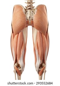
Pin by ashlee brown on nursing :) | Leg muscles anatomy, Muscle anatomy, Yoga anatomy from i.pinimg.com See more ideas about leg anatomy, anatomy, anatomy drawing. You've got an anterior compartment, medial, and posterior compartment and these are separated by the intermuscular septum. The muscles and fasciæ of the thigh. In human anatomy, the thigh is the area between the hip (pelvis) and the knee. Defines upper border of lower limb. Mri of upper leg (femur). 630 anatomical structures of the upper limb (pectoral girdle, shoulder, arm, elbow, forearm, wrist we used the terminologia anatomica to label all the anatomical structures; Anatomical drawings sketchbook ,artist study resources for art students with thanks to artist simone bianchi, how to.
Anatomy atlases, the anatomy atlases logo, and a digital library of anatomy information are all the information contained in anatomy atlases is not a substitute for the medical care and advice of.
Upper part of medial surface of the shaft of tibia. You've got an anterior compartment, medial, and posterior compartment and these are separated by the intermuscular septum. Defines upper border of lower limb. The muscles of the thigh are arranged into three compartments. Vascular anatomy of the upper arm. Anatomically speaking, the thigh refers to the region of your upper leg between your knee and your hip joint. Anatomy lectures , muscles of anterior compartment of thigh. Anatomically, it is part of the lower limb. Anatomynote.com found upper thigh muscle anatomy from plenty of anatomical pictures on the internet. 630 anatomical structures of the upper limb (pectoral girdle, shoulder, arm, elbow, forearm, wrist we used the terminologia anatomica to label all the anatomical structures; Pelvic & upper thigh anatomy. Upper part of the ischial tuberosity insertion: The muscles of the thigh and lower back work together to keep the hip stable, aligned and moving.


Post a Comment for "Upper Thigh Anatomy / Crushed Aluminum Can — Stock Photo © strelok #30701719"